Advances in Vet Imaging Tech
Veterinary medicine has made tremendous strides in recent years, particularly in the field of imaging technology. Advances in imaging tech have made it possible for vets to diagnose and treat a wider range of diseases, as well as gain a better understanding of the health of animals.
As the pet healthcare industry continues to evolve, veterinary imaging technology is becoming increasingly advanced to provide the most accurate and precise information possible. Recent advances in veterinary imaging have resulted in improved image quality, lower radiation doses, faster acquisition times, and a wider variety of applications.
The advent of modern veterinary imaging technologies has revolutionised the field of veterinary medicine, offering unprecedented insight into the physical and physiological processes occurring in animals. These technologies, including radiography, ultrasound, computed tomography (CT), magnetic resonance imaging (MRI), and positron emission tomography (PET), enable veterinarians to diagnose a wide range of conditions more accurately and quickly than ever before.

New Possibilities for Pet Scans
The advent of pet vet scans has opened a multitude of possibilities in the field of veterinary medicine. Through innovative imaging technology, veterinarians now have the opportunity to non-invasively evaluate and diagnose a wide array of conditions and diseases in companion animals. This breakthrough has allowed for early detection of serious illnesses and improved treatment outcomes. In addition, these scans are applicable for the evaluation of musculoskeletal conditions, providing detailed insights into potential abnormalities that may have previously been overlooked.
The scans are a novel technological advancement that provides unique advantages for both pet owners and veterinarians. This non-invasive imaging technique offers a number of benefits over traditional radiography, including enhanced visualisation of soft tissue structures, reduced radiation exposure to both the patient and operator, and improved diagnostic accuracy. The use of pet vet scans also offers a degree of flexibility with regard to patient positioning, providing improved visualisation for multiple views in a single session.
Decoding Veterinary X-Rays
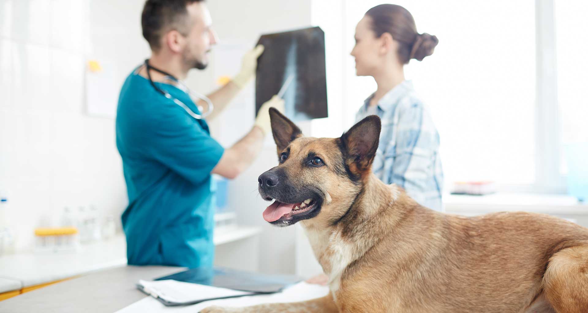
Decoding veterinary x-rays is a process of utilising specialised imaging equipment to acquire a radiographic representation of an animal’s internal anatomy. As part of the process, the images are then evaluated by an experienced and trained professional who is capable of interpreting the radiographs in order to diagnose any underlying medical conditions or diseases. This diagnostic information is then used to develop a personalised treatment plan for the animal, specifically tailored to its individual needs and condition.
Veterinary X-Rays offer a variety of advantages, most notably the ability to identify and diagnose an array of pathologies. Such diagnostics are critical in order to ascertain the necessary course of treatment and to provide a more accurate prognosis. Furthermore, X-Rays can be used to monitor the progression or regression of certain pathologies as well as providing additional information through specialised techniques such as computed tomography scanning.
How Does Veterinary Medical Imaging work?
Veterinary medical imaging is a fundamental practice utilised in veterinary medicine. It uses sophisticated technology to produce visual representations of the anatomy and physiology of an animal, enabling the identification of diseases and other abnormalities. The most common types of veterinary medical imaging include radiographs, ultrasonography, computed tomography (CT), magnetic resonance imaging (MRI), and nuclear scintigraphy.
Each modality has its own advantages and limitations in terms of image resolution, cost effectiveness, and availability. This can include conventional radiography, ultrasonography, computed tomography (CT), magnetic resonance imaging (MRI), nuclear medicine, and positron emission tomography (PET). It is an invaluable tool for veterinarians as it allows for accurate diagnosis and effective treatment planning.
Exploring the Benefits of Veterinary Medical Imaging
The application of veterinary medical imaging techniques has been seen to provide a wealth of benefits in the field of animal healthcare. Utilising technologies such as radiography, ultrasound, computed tomography, and magnetic resonance imaging, practitioners are able to diagnose a range of conditions with heightened accuracy and precision. Moreover, through the use of these modalities, practitioners are able to gain a better understanding of the extent and complexity of disease processes which may be present without needing to resort to exploratory surgery.
The advancements in veterinary imaging technology have greatly improved the accuracy and speed with which veterinarians can diagnose and treat animals. These technologies provide more detailed images than ever before, allowing for greater insight into an animal’s health. Additionally, these technologies are much more affordable than they were in the past, so an increasingly larger number of people are able to access them. As a result, this technology is becoming a standard part of everyday life in the veterinary world as it continues to improve and evolve.
